
Figure 10.
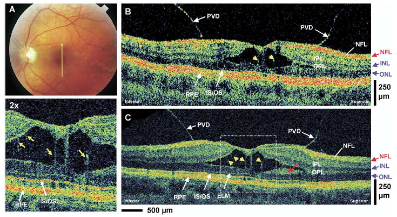
Patient 8. A, Fundus photograph depicting vitreomacular traction and the direction of optical coherence tomography (OCT) scans. B, C, StratusOCT and ultrahigh-resolution (UHR) OCT images, respectively, of a patient with vitreomacular traction. Posterior vitreous detachment (PVD) can clearly be visualized in both images. The enlargement of the UHR OCT image shows several distended structures (yellow arrows) spanning the separation of the sensory retina that are suggestive of Müller cells. A highly backscattering layer in the UHR OCT image that is adjacent to the outer nuclear layer (ONL) (red arrow) might be a portion of the outer plexiform layer (OPL). ELM = external limiting membrane; INL = inner nuclear layer; IPL = inner plexiform layer; IS/OS = junction between the photoreceptor inner segment and outer segment; NFL = nerve fiber layer.