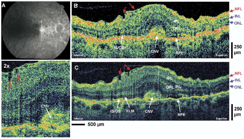
Figure 7.

Patient 5. A, Late-phase fluorescein angiogram depicting choroidal neovascularization (CNV) and the direction of optical coherence tomography (OCT) scans. B, C, StratusOCT and ultrahigh-resolution (UHR) OCT images, respectively, of a patient with CNV. Small irregularities in the nerve fiber layer (NFL) and ganglion cell layer with evidence of epiretinal membrane formation (red arrows) can be observed in the UHR OCT image and its enlargement. ELM = external limiting membrane; INL = inner nuclear layer; IPL = inner plexiform layer; IS/OS = junction between the photoreceptor inner segment and outer segment; ONL = outer nuclear layer; OPL = outer plexiform layer.
