
Figure 9.
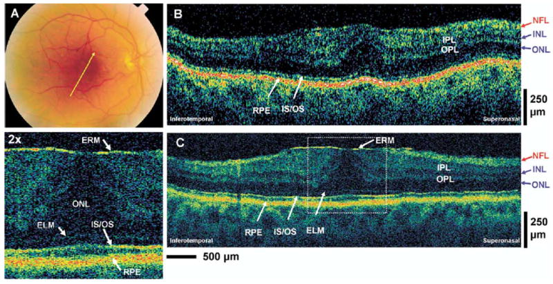
Patient 7. A, Fundus photograph depicting the epiretinal membrane (ERM) and the direction of optical coherence tomography (OCT) scans. B, C, StratusOCT and ultrahigh-resolution (UHR) OCT images, respectively, of a patient with an ERM. The enlargement of the UHR OCT image clearly shows the thin, highly backreflecting epiretinal membrane that is not visualized in the StratusOCT image. ELM = external limiting membrane; INL = inner nuclear layer; IPL = inner plexiform layer; IS/OS = junction between the photoreceptor inner segment and outer segment; NFL = nerve fiber layer; ONL = outer nuclear layer; OPL = outer plexiform layer; RPE = retinal pigment epithelium.