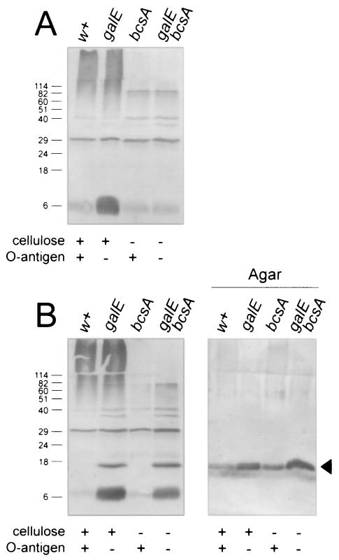
FIG. 2.
Western blot analysis of S. enterica serovar Enteritidis 3b ΔagfB donor and ΔagfA recipient strains for the production of AgfA. S. enterica serovar Enteritidis 3b ΔagfA (A) and ΔagfB (B) strains were analyzed for production of AgfA after growth on T agar. Samples from strains were loaded as indicated, and strain phenotypes are listed below each lane. Panel A and the first four lanes of panel B represent proteins in the debris left over after boiling cells in SDS-PAGE sample buffer, whereas agar samples (B) represent proteins present in agar plugs after cells were removed. All samples were treated with formic acid prior to loading on SDS-PAGE. AgfA and associated material were detected by using immune serum raised to purified Tafi; the arrowhead in panel B indicates monomeric AgfA. Molecular mass markers (in kilodaltons) are indicated on the left.

