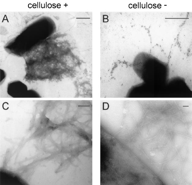
FIG. 4.
Transmission electron microscopy of Tafi fimbriae. Fimbriae produced by S. enterica serovar Enteritidis strain 3b (cellulose positive) (A and C) or the ΔbcsA mutant (cellulose negative) (B and D) were immunogold labeled with AgfA-specific monoclonal antibody 3A-12 ascites followed by goat anti-mouse immunoglobulin-10-nm-diameter gold (A and B) or simply negatively stained with uranyl acetate (C and D). Bars, 500 nm (A and B) or 100 nm (C and D).

