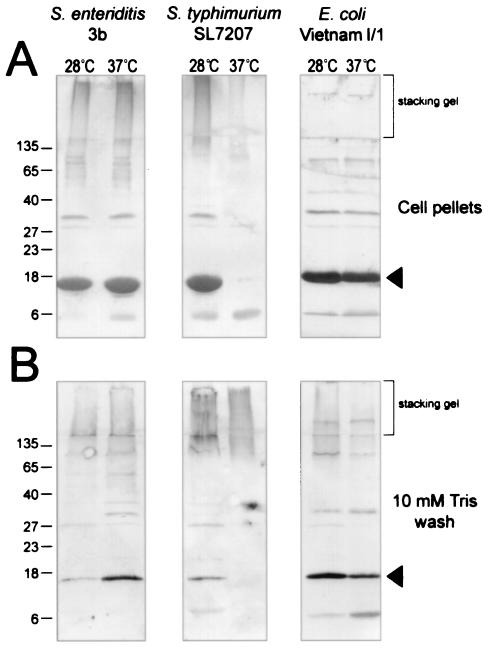
FIG. 5.
Detection of immunoreactive Tafi-associated material in different enterobacterial species. Proteins in the debris left over after boiling cells in SDS-PAGE sample buffer (cell pellets) or acetone-precipitated proteins from a 10 mM Tris (pH 8) wash of whole cells of S. enterica serovar Enteritidis 3b, S. enterica serovar Typhimurium SL7207 or E. coli Vietnam I/1 after growth on T agar at 28 or 37°C were loaded as indicated. The brackets show the regions of immunoblots corresponding to the stacking gel from SDS-PAGE. AgfA and associated material were detected by using immune serum raised to whole Tafi; arrowheads indicate monomeric AgfA (Salmonella) or CsgA (E. coli). Molecular mass markers (in kilodaltons) are indicated on the left.

