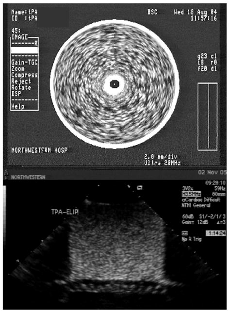
Figure 1.

Top: Image of tPA-loaded ELIP in a glass vial imaged with a 20 MHz intravascular ultrasound catheter (25× dilution). The central dark spot corresponds to the imaging catheter. Bottom: Image of tPA-loaded ELIP in an imaging well imaged with a 3.5 MHz harmonic transthoracic probe (100× dilution). The artifact at the bottom of the image corresponds to the Rhocee rubber used as sound-absorbing material.
