
Fig. 4.
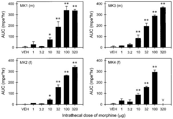
Different doses of intrathecal morphine-induced antinociception against 50°C water during the six test sessions in four monkeys. Abscissae (all panels): different doses (micrograms) of intrathecal morphine. Ordinates (all panels): area under curve (percent maximum possible effect × hours). Asterisks represent a significant difference (*P < 0.05; **P < 0.01) from the vehicle (VEH) condition. Other details are as in figures 2 and 3.