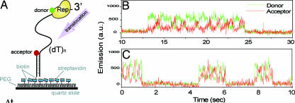
Fig. 2.
Explorations of the sliding behavior of Rep on a single-stranded DNA segment attached to a surface. (A) Schematic of the labeling arrangement for FRET measurements. Traces (B, 22°C; C, 37°C) of donor (green) and acceptor (red) fluorescence signals for a single Rep molecule are shown. [Reproduced with permission from ref. 25 (Copyright 2005, MacMillian Publishers, Ltd.).]

