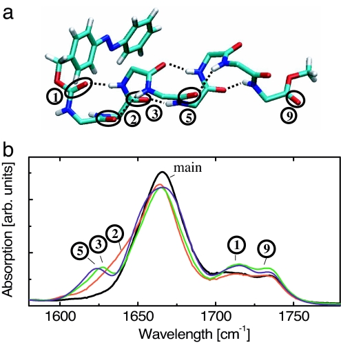
Fig. 1.
Structure and steady-state spectroscopy. (a) X-ray structure of PAZ-Aib-Ala-(Aib)6-OMe (molecule A, only the backbone is shown). The tags 1 to 9 refer to the corresponding absorption bands in b and count C O groups with increasing distance from the azobenzene moiety. (b) FTIR absorption spectra of the 13C-unlabeled reference molecule (PAZ-Aib-Ala-Aib6-OMe, black line), and those of the 13C-labeled peptides PAZ-Ala*-Aib7-OMe (red), PAZ-Aib-Ala*-Aib6-OMe (green), and PAZ-Aib3-Ala*-Aib4-OMe (blue).

