Fig. 1.
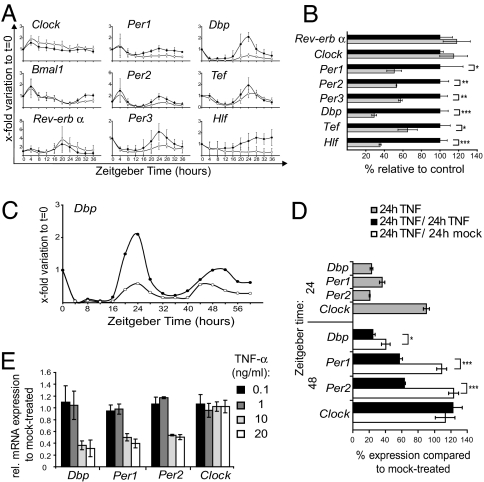
TNF-α impairs expression of clock genes in synchronized NIH 3T3 fibroblasts. (A) TNF-α attenuates circadian Per1/2/3 and PAR bZIP family (Dbp, Tef, and Hlf) gene expression. After serum shock (ZT 0–2), cells were kept in serum-free medium with TNF-α (10 ng/ml; open circles) or without the cytokine (filled circles) and analyzed every 4 h with quantitative real-time RT-PCR. Results are shown as x-fold variations to nonsynchronized fibroblast cultures at ZT 0; three independent experiments; mean values ± SD. (B) Per and PAR bZIP family genes are significantly down-regulated at the 24-h peak (TNF-α: gray bars; controls: black bars), whereas Clock and RevErbα are not affected. Expression of Rev-Erbα was assessed at its peak at ZT 20. Data show one representative experiment done in triplicate (mean ± SD) of four experiments. (C) The amplitude of Dbp expression in serum-shocked NIH 3T3 fibroblasts was attenuated during 60 h in the presence of TNF-α (open circles) compared with controls (filled circles). (D) Withdrawal of TNF-α after the first peak at ZT 24 shows that the suppression of Dbp, Per1, and Per2 at the second peak at ZT 48 is reversible. After serum shock, cells were treated with TNF-α for 24 h (gray bars), or for 48 h with or without a withdrawal of TNF-α after 24 h (white and black bars, respectively). Gene expression was compared with mock-treated cells at the respective ZT (100% expression). Data show one representative experiment done in triplicates (mean ± SD) of three experiments. (E) The suppression of the expression of Dbp, Per1, and Per2 in synchronized fibroblasts at ZT 24 is dose-dependent being significant (P < 0.005) at doses higher than 1 ng/ml TNF-α. Data show the mean ± SD of three independent experiments performed in triplicates. For BDE, we used the independent-sample t test; *, P ≤ 0,05; **, P ≤ 0,005; ***, P ≤ 0.0005.

