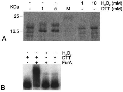
FIG. 3.
Electrophoretic mobility of the FurA protein and binding of FurA to DNA under different redox conditions. (A) Purified M. tuberculosis FurA (1.7 μg) expressed in E. coli was run in the first lane of an SDS-15% polyacrylamide gel. Equivalent samples were mixed with either DTT or H2O2 at the concentrations indicated above the lanes before being loaded on the gel. Lane M, molecular mass markers, with sizes indicated on the left. (B) Gel shift caused by FurA. The DNA probe covers the −154/+33 M. tuberculosis region. FurA (10 μM) was added, as indicated above the lanes, to 32P-labeled DNA probe in the presence of 200 μM Ni2+, and complexes were resolved on a Tris-acetate-8% polyacrylamide gel containing 200 μM Ni2+. DTT (1 mM) and H2O2 (10 mM) were added to the incubation buffer as indicated.

