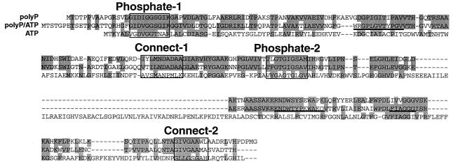
FIG. 2.
Comparison of the deduced amino acid sequences of the M. phosphovorus polyP-GK, the M. tuberculosis polyP/ATP-GK, and the E. coli ATP-GK. The shaded amino acids were conserved among the GKs (CLUSTALW analysis; DDBJ). The Phosphate-1, Phosphate-2, Connect-1, and Connect-2 motifs are boxed. The underlined sequences (WRGPLGVTYPGV, KNDWTYPKWAKQ, and FIAGGG) are the proposed regions of glucose, polyP, and adenosine binding in the polyP/ATP-GK (9), respectively.

