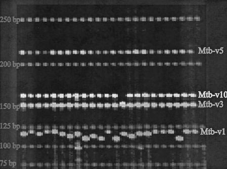
FIG. 2.
Electrophoresis of MLVA fragments. Fluorescent image of markers Mtb-v1,-v3,-v5, and -v10 on an ABI377 electrophoresis gel. Each marker allele is identified through a unique size and color combination, allowing adequate discrepant ability across comparable-sized fragments from sundry alleles. Actual sizes of these alleles can be seen in Table 4. Sizes are shown in nucleotide bases.

