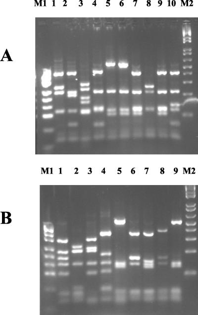
FIG. 2.
fliC RFLP patterns generated using MspI (A) and HaeIII (B). All patterns observed in this study are shown. Lane 4 in panel A and lane 5 in panel B are RFLP patterns indicative of fliC RFLP group I. Lanes 5 and 6 in panel A are from two isolates with the same MspI RFLP pattern. M1, pUC19/MspI (Helena Biosciences); M2, 1-kb ladder (Helena Biosciences).

