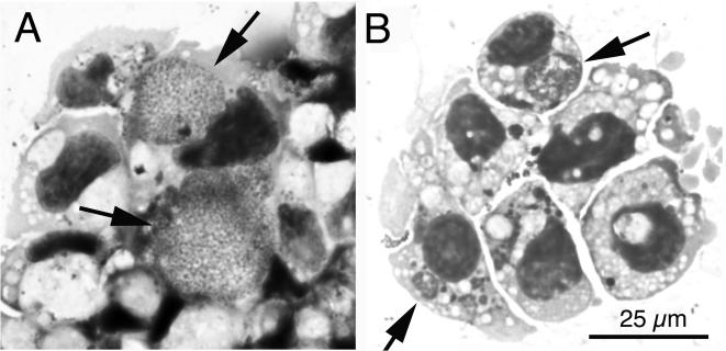
FIG. 1.
Light microscopic appearance of the WTD-Anaplasma isolate in Giemsa-stained cytocentrifuge preparations of tick cell culture. (A) WTD-Anaplasma isolate from WDT76 in its eighth passage in tick cells. Arrows indicate large intracytoplasmic inclusions filled with numerous bacteria. (B) WTD-Anaplasma reisolated from WTD86. Arrows point to intracytoplasmic morulae. The scale bar in panel B gives the magnification for both panels.

