FIG. 2.
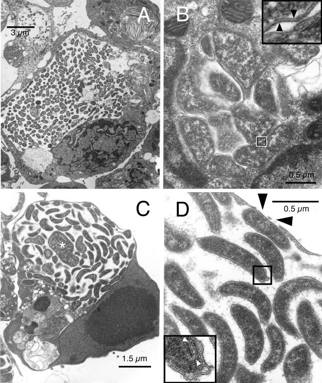
Ultrastructure of the WTD-Anaplasma in tick cell culture. (A) Inclusion that appears to occupy most of its host cell. Note the lack of association of the bacteria with the endosomal membrane. (B) Image of a small morula with pleomorphic bacteria that tightly abut each other and the endosomal membrane. Inset, high magnification of the boxed-in area, showing the double-layered profile of the cell wall (arrowheads). (C) Third type of inclusion containing primarily slightly curved rods but also larger and more irregular organisms (asterisk). Note the intimate association of Anaplasma with the endosomal membrane. (D) Higher magnification of panel C. Black arrowheads indicate gaps in the endosomal membrane; white arrowheads within the inset point to folded membrane structures at one end of a curved rod, enlarged from the boxed-in area.

