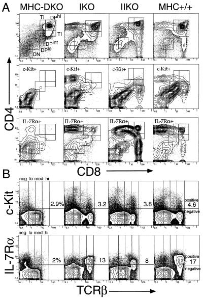
Figure 1.
Distribution of c-Kit+ or IL-7Rα+ cells in either MHC-DKO, MHC-IKO, MHC-IIKO, or MHC+/+ thymus. (A) The CD4/CD8 profiles of total thymocytes (Top), those of c-Kit+ cells (Middle), and those of IL-7Rα+ cells (Bottom). Squares indicate the definition of DN, DPlo, DPint, DPhi, and TI (CD4+CD8med or CD4medCD8+) stages in this study. (B) The TCRβ/c-Kit (Upper) and the TCRβ/IL-7R (Lower) profiles of total thymocytes. Note that MHC-DKO mice lack the TCRmed-hi c-Kit+ and TCRmed-hi IL-7R+ populations.

