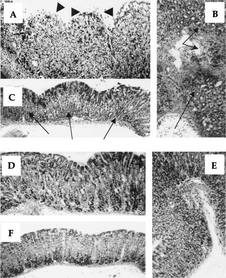
FIG. 4.
Photomicrographs of hematoxylin-eosin-stained gastric tissues of SPF (A, B, and C) and GF (D, E, and F) mice prevaccinated with H. pylori HSP60 at 10 weeks p.i. Cardia mucosa of SPF mice frequently showed severe gastritis with disruption of the gland structure, severe inflammatory cell infiltration, and erosion (A). GF mice did not show these symptoms (D). Severe inflammatory cell infiltration was observed in mucosa in SPF mice (B) but not in GF mice (E). Antral mucosa of SPF mice also showed severe inflammatory cell infiltration (C), but GF mice did not (F). Arrows indicate inflammatory cell infiltration, and arrowheads indicate erosion. Magnification, ×200.

