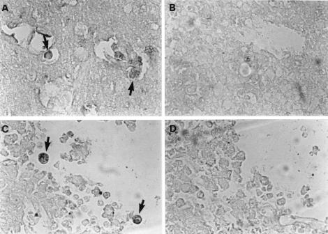
FIG. 4.
Mouse brain tissue infected with N. fowleri trophozoites was treated with an anti-Nfa1 antibody (A and C) and unimmunized control serum (B and D). Trophozoites (arrows) were stained brown. (A and B) The inflammatory region of the brain tissue; (C and D) the necrotic region of the brain tissue. Magnification, ×200.

