
Figure 1.
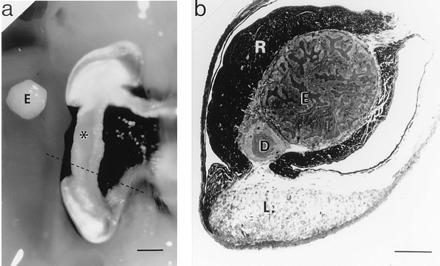
The symbiotic organ of E. scolopes. (a) Ventral view of the right lobe of the organ. Alongside of this lobe is placed the bacteria-containing epithelial tissue (E), which has been dissected out the left lobe of the organ. ∗, Position of this tissue in the right lobe. The dashed line indicates the location of the cross section shown in b. (Bar = 1 mm.) (b) Light micrograph of a cross section of an E. scolopes symbiotic organ. The section was stained with Richardson’s stain (8). E, bacteria-containing tissue comprised of epithelial cells surrounding crypts that house V. fischeri; D, ciliated duct; R, reflector tissue; L, light organ lens. (Bar = 450 μm.)