
Figure 6.
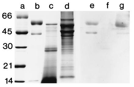
SDS/PAGE and immunoblot, using anti-FPO, of squid tissues and purified human MPO. For immunoblots, the soluble proteins were prepared as described in Fig. 3, except that they were homogenized in a 50 mM sodium phosphate buffer with 0.1 M NaCl (pH 7.2). SDS/PAGE was performed as described in Fig. 3. Proteins were electrophoretically transferred from unstained gels onto nitrocellulose membrane (modified from ref. 26). Immunoblots were performed with a chemiluminescence detection system (Renaissance Kit; DuPont/NEN). Membranes were first blocked for 12 h at 4°C in 50 mM Tris, 150 mM NaCl, 0.5% Tween 20 (pH 7.5) (TTBS) containing 3% powdered milk and a 1:100 dilution of goat serum. Following this, they were incubated in a 1:100 dilution of anti-FPO in TTBS for 12 h at 4°C and subsequently incubated in the secondary antibody, goat anti-rabbit IgG conjugated to horseradish peroxidase. Preimmune serum, obtained from the rabbit before immunization with FPO, was substituted for the 1° antiserum as a negative control. Lane: a, standards; b and e, purified human MPO (0.5 μg each lane); c and f, squid digestive gland (20 μg each lane); and d and g, bacteria-containing epithelial tissue of the symbiotic organ (20 μg each lane). The molecular masses of the standards are shown in kDa.