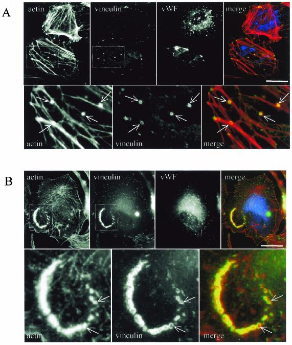
FIG. 10.
Podosomes are detected on aortic endothelial cells in a primary culture. Human endothelial cells were prepared from freshly explanted aorta fragments and directly seeded on glass coverslips (A) or on fibronectin-coated coverslips (B). Cells were fixed and processed for immunofluorescence with rhodamine-phalloidin, anti-vinculin, and anti-Von-Willebrand factor antibodies (vWF). Note the presence of podosomes (arrows) visualized as actin dots surrounded by vinculin rings. Bar, 50 μm.

