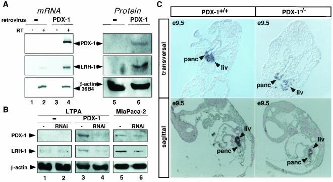
FIG. 6.
PDX-1 regulates the expression of LRH-1 in vivo. (A) Expression of PDX-1 and LRH-1 analyzed by RT-PCR (mRNA) and immunoblotting (protein) in LTPA cells overexpressing PDX-1 (PDX-1) or not (−) by retroviral infection. Reverse transcription reactions were performed with (RT+) or without (RT−, negative control) reverse transcriptase. Subsequent PCR was performed with primers specific for PDX-1, LRH-1, and 36B4. Immunoblotting was performed with specific antibodies raised against PDX-1, LRH-1, and β-actin. The intensity of the signals was quantitated by phosphorimager analysis, and the induction was determined after normalization to β-actin signals. (B) Decreasing PDX-1 levels by RNAi reduces LRH-1 expression. Immunoblotting of protein isolated from control LTPA cells (−), LTPA cells retrovirally overexpressing PDX-1 (PDX-1), or MiaPaca-2 cells, all transfected with an empty RNAi vector (−, lanes 1, 3, and 5) or an RNAi vector targeting the PDX-1 mRNA (RNAi, lanes 2, 4, and 6). The induction was calculated as described for panel A. (C) Expression of LRH-1 in transversal (upper panel) and sagittal (lower panel) sections of PDX-1+/+ and PDX-1−/− mouse embryos from stage E9.5 analyzed by in situ hybridization. LRH-1 expression was significantly stronger in pancreas (panc) and liver (liv) of 2 PDX-1+/+ relative to PDX-1−/− tissues.

