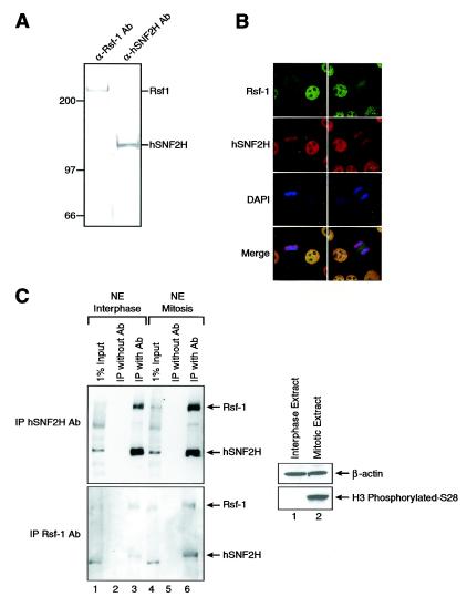
FIG. 2.
Immunofluorescence microscopy of RSF. (A) Western blots with RSF antibodies. Partially purified fractions of RSF derived from HeLa nuclear pellet were Western blotted with monoclonal antibodies against the RSF subunits Rsf-1 and hSNF2H. (B) Immunofluorescence microscopy. HeLa cells were fixed and permeabilized, followed by incubation with antibodies (Ab) against the RSF subunits Rsf-1 and hSNF2H. The antibody against Rsf-1 is shown in green, the antibody against hSNF2H is shown in red, and DAPI (4′,6′-diamidino-2-phenylindole) is shown in blue. The bottom panel shows the merge between all of them. Two cells are shown on the field. The cell to the left is in mitosis (metaphase), whereas the cell to the right is in interphase. (C) Interaction between Rsf-1 and hSNF2H during mitosis. Western blot analysis of immunoprecipitation analysis carried out with hSNF2H (top panel) and Rsf-1 (bottom panel) antibodies, as indicated in the left side of the figure. The input for the immunoprecipitation (IP) is indicated on top of the figure: interphasic (left side) or mitotic (right side) nuclear extract. As a negative control, the extract was incubated only with protein G-agarose and processed like the other samples. We used antibodies against histone H3 and phosphorylated S28 as a marker for mitosis and β-actin as a loading marker.

