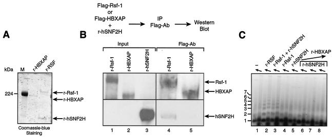
FIG. 6.
Comparison of Rsf-1 and HBXAP. (A) Coomassie blue staining of recombinant RSF obtained by coexpression of the subunits and HBXAP. The arrow indicates the migration position of the RSF subunits Rsf-1 and hSNF2H and of HBXAP. M, molecular marker. (B) Interaction between hSNF2H and HBXAP. Western blot analysis of the immunoprecipitation carried out as outlined at the top of the figure. Flag-tagged HBXAP or Flag-tagged Rsf-1 was incubated with hSNF2H and immunoprecipitated with antibody against Flag. The input shows the starting material for the immunoprecipitation before the subunits were mixed. (C) Chromatin assembly. An agarose gel of a micrococcal nuclease digestion of a chromatin assembly reaction carried out with HBXAP, as follows: without factor (lane 1), with recombinant RSF obtained by coexpression of the subunits (lane 2), with recombinant Rsf-1 and hSNF2H together (lane 3), with recombinant Rsf-1 alone (lane 4), with recombinant hSNF2H alone (lane 5), and with recombinant hSNF2H plus increasing amounts of HBXAP (between 0.16 and 1 μg) (lanes 6 to 8).

