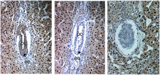
FIG. 9.
Immunohistological analysis with Csn8 antibody. (A) A 6.5 dpc normal embryo. (B and C) Mutants at 6.5 dpc (B) and 7.5 dpc (C). Panels A and B show embryos from the same litter. Panel C and panel H in Fig. 5 show different sections of the same embryo. ee, embryonic ectoderm (or epiblast cells); pc, proamniotic cavity. Bars, 100 μm.

