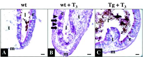
FIG. 2.
Transgenic expression of dnTR inhibits T3-induced intestinal remodeling. Wild-type (wt) or transgenic (Tg) tadpoles were treated with T3 as in Fig. 1. Cryosections of isolated intestines were stained with methyl green pyronine Y. (A) Note that the untreated control intestine had thin muscle layers (m) and sparse connective tissue except in the typhlosole (t), which is the single epithelial fold occupying much of the lumen (l) in the larval intestine (only part of the typhlosole is shown in the image). The majority of staining was seen in the larval epithelium (e). (B) In wild-type tadpoles after 3 days of T3 treatment, the muscle and connective-tissue layers increased in thickness largely due to active cell proliferation (56). The labeling in the larval epithelial cells decreased dramatically as these cells underwent apoptosis (arrows). Adult epithelial cells (arrowheads), whose origin remains unknown, were induced to proliferate and show intense staining. (C) All of these changes were prevented in the transgenic tadpoles treated with T3 for 3 days. Note that the typlosoles differ in size because of intestinal remodeling and/or the sections were from different locations within the anterior intestine. The brown material visible in panels A and C is gut contents in the lumen. The results are representative of three transgenic tadpoles examined. Bar, 20 μm.

