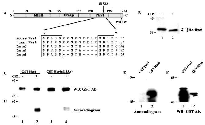
FIG. 9.
Analysis of Hes6 phosphorylation. (A) Schematic representation of the domain structure of Hes6. Indicated are the bHLH domain, the Orange domain predicted to form helices 3 and 4, the PEST region containing the SPXSS-SDXE motif and its resident Ser183, and the WRPW tetrapeptide. Shown in detail are the sequences of the SPXSS-SDXE elements from mouse and human Hes6 (3) and Drosophila Enhancer of split m5, m7, and m8 (37). Invariant residues are indicate in boldface. (B) 293A cells were transfected with HA-Hes6, and cell lysates were incubated in the absence or presence of calf intestinal phosphatase (CIP), followed by Western blotting with anti-HA antibodies. (C and D) The indicated GST fusion proteins were purified and subjected to in vitro phosphorylation in the absence (lanes 1 and 3) or presence (lanes 2 and 4) of purified protein kinase CK2, followed by autoradiography (D) and Western blotting (WB) with anti-GST antibodies (Ab.) (C). (E and F) The indicated GST fusion proteins were purified and subjected to in vitro phosphorylation in the presence of purified protein kinase CK2, followed by autoradiography (E) and Western blotting with anti-GST antibodies (F). Positions of size standards are indicated in kilodaltons in panels B to F.

