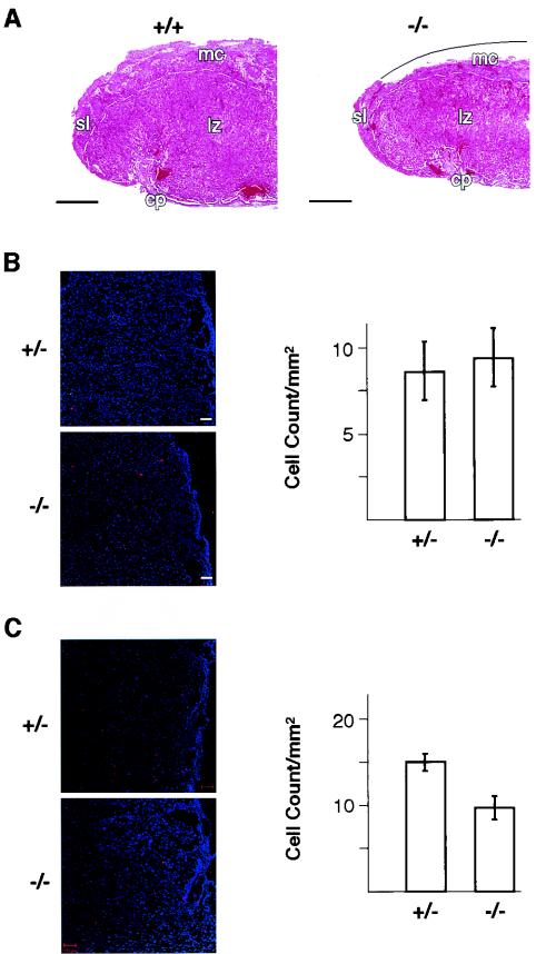
FIG. 4.
Analysis of the placentas of Plk2−/− embryos. (A) Hematoxylin- and eosin-stained section of half of a placenta showing diminished labyrinthine zone, indicated by white dashed lines, in Plk2−/− (−/−) embryos. The curved solid line depicts a portion of the maternal component that was peeled off during sample preparation. mc, maternal (decidual) component; sl, spongiotrophoblast layer at periphery of the placenta; lz, labyrinthine zone; cp, chorionic plate. +/+, Plk2+/+. Bar, 0.5 mm. (B) The labyrinthine zones of Plk2+/− (+/−) and Plk2−/− embryos contain similar numbers of apoptotic cells. On the left are fluorescence confocal microscopy images of TUNEL-stained (red) placenta sections. Cell nuclei were stained with DAPI (4′,6′-diamidino-2-phenylindole) (blue). On the right, counts of TUNEL-stained cells from five samples are summarized. The error bars indicate standard deviations. (C) Phospho-histone H3 staining shows that fewer cells are proliferating in the labyrinthine zone of the placenta of Plk2−/− embryos than in Plk2+/− embryos. On the left, fluoresence confocal microscopy images of sections of the labyrinthine zone are shown. Phospho-histone H3-positive cells are stained red, and cell nuclei are blue. On the right, counts of phospho-histone H3-positive cells from five tissue sections are summarized. The results are representative of two independent experiments.

