FIG. 1.
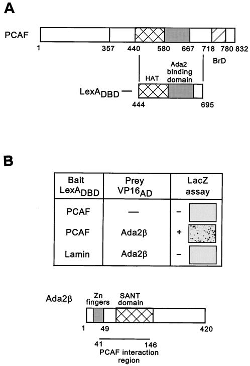
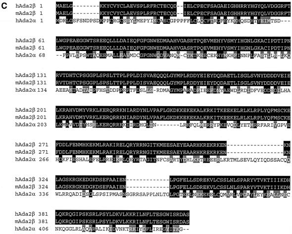
LexADBD-PCAF two-hybrid screen. (A) PCAF domain structure. Nucleosomal recognition, aa 1 to 357; HAT domain, aa 440 to 580; Ada2-binding domain, aa 580 to 667; bromodomain, aa 718 to 780. The region of PCAF (aa 444 to 695) used as bait in the two-hybrid screen is shown below. (B) Two-hybrid interaction between PCAF and Ada2β. In the upper part of the panel, LexADBD-PCAF444-695 or LexADBD-Lamin were cotransformed into yeast with the amino terminus of Ada2β fused to the VP16 activation domain. Interaction was tested by using a LacZ assay of colonies transferred to filters. Positively interacting blue colonies are shown after 30 min of color development at 30°C. In the lower part of the panel is a diagram of Ada2β. Domains conserved between different Ada2 species are indicated. The region of interaction with PCAF is indicated. (C) Sequence alignment of hAda2β, mAda2β, and hAda2α. Alignment was performed by using CLUSTAL W. Identical amino acids are shown in black boxes, and similar amino acids are shown in gray boxes.Continued.


