
Fig. 4.
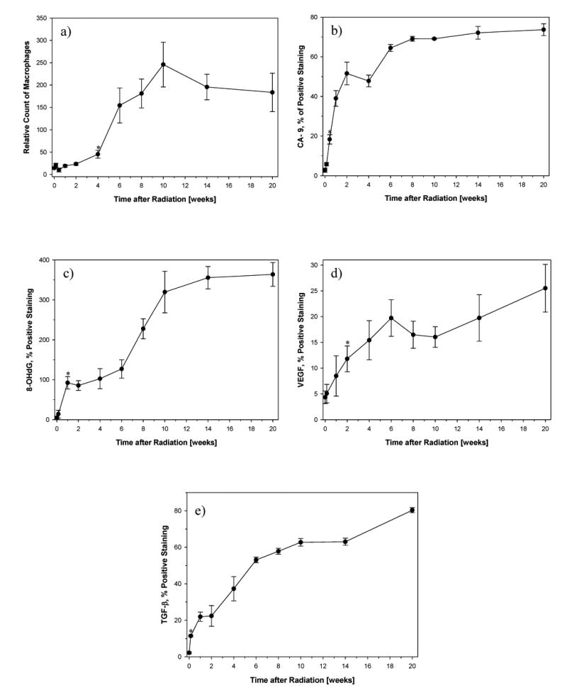
Quantitative analyses from immunohistochemistry images prior to and at different times after irradiation of the right hemithorax with a single dose of 28 Gy. Data are represented as mean obtained from 5 animals per timepoint, error bars are ± 1 SEM. *p<0.05, first significant change compared with no radiation.
- Number of activated macrophages (ED-1): A significant increase in macrophages was seen 4 weeks after irradiation (p=0.0231) and peaked at 10 weeks (p=0.00979).
- Hypoxia (CA-9): A significant increase in CA-9 expression was already seen 3 days after irradiation (p=0.000327) and increased further with time.
- ROS (8-OHdG): A significant increase in 8-OHdG expression started 1 week after irradiation (p=0.0097) and increased further with time.
- VEGF: A significant increase in VEGF expression started 2 weeks after irradiation (p=0.0284).
- TGF-β: A significant increase in TGF-β expression was already seen 1 day after irradiation (p=0.00146) and continuously increased further with time.