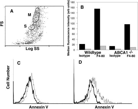
Figure 6. Identification and annexin V staining of macrophages in peritoneal lavage from wildtype or ABCA1−/− mice.
Cells from peritoneal lavage were stained with mAb F4/80 (or isotype control mAb) or with annexin V on ice and examined by flow cytometry at room temperature. A, forward (FS) vs side (SS) light scatter plot, with cells having typical macrophage light scatter characteristics in the M gate and Asmall@ cells in the S gate. B, median fluorescence intensity of cells in the M gate (black) or S gate (grey) following staining with mAb F4/80 or isotype control mAb. C&D, annexin V staining, in the presence (thin line) or absence (thick line) of Ca2+, of cells in the M gate from wildtype (C) or ABCA1−/− (D) mice.

