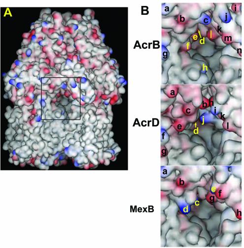
FIG. 6.
Vestibules of AcrB, AcrD, and MexB. (A) The trimeric AcrB pump is viewed from the outside, so that the two protomers are seen in front and the third protomer is mostly hidden in the back. The vestibule, at the interface of the two protomers, is indicated by the rectangle in the center. Acidic residues are colored red, and the basic residues are in blue. (B) The central region of the view shown in panel A, containing the area surrounding the vestibule, is shown in a larger magnification. Charged amino acid residues on the surface are identified by letters. For AcrB, Lys322 (a), Glu314 (b), Lys312 (c), Asp101 (d), Asp99 (e), Asp301 (f), Lys334 (g), Lys29 (h), Glu705 (i), Lys850 (j), Asp99 (k) (from the protomer on the right), Glu95 (l), Glu842 (m), and Glu839 (n) are indicated. For AcrD, Glu322 (a), Glu312 (b), Asp311 (c), Asp99 (d), Glu304 (e), Lys334 (f), Glu338 (g), Glu856 (h), Lys847 (i), Lys95 (j), Glu843 (k), and Asp840 (l) are shown. For MexB, Lys322 (a), Glu314 (b), Lys170 (c), Lys304 (d), Lys848 (e), Glu845 (f), Glu95 (g), and Asp838 (h) are indicated. The hypothetical structures of AcrD and MexB were generated by the SWISS-MODEL program (http://www.expasy.ch) by using the crystal structure of AcrB as the template. The drawing was made with the program DS Viewer Pro 5.0 (Accelrys, San Diego, Calif.).

