
Figure 6.
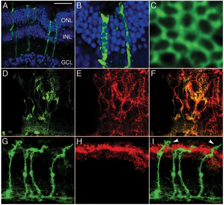
LV vector delivered eGFP expression in healthy and diseased retinas. One month after VSV-CD44-GFP vector injection at P21 (A) eGFP-positive Müller cells are observed spanning the entire thickness of SD rat retina (A–F) far from the injection site (ONL curvature is a tissue processing artifact). (B) Müller cell processes surround DAPI-stained photoreceptor nuclei (blue). (C) High-magnification en face view of Müller cell fiber basket matrix at the OLM. (D) eGFP positive, (E) glutamine synthetase–stained (red), and (F) merged Müller cells are disorganized, most likely as a result of the subretinal injection procedure. Two months after VSV-GFAP-GFP vector injection at P21, (G) eGFP-positive Müller cells were observed in the diseased S334Ter± retina (G–I). (H) Photoreceptor outer segments were stained with a rhodopsin antibody (red). (I) The merged image indicates the relationship between the Müller cells and the photoreceptor outer segments; Müller cell apical processes (arrowheads) have emerged beyond the OLM and into the subretinal space. DAPI (blue) was used to counterstain nuclei in (A–B). Scale bar, 50 μm.