
Fig. 2.
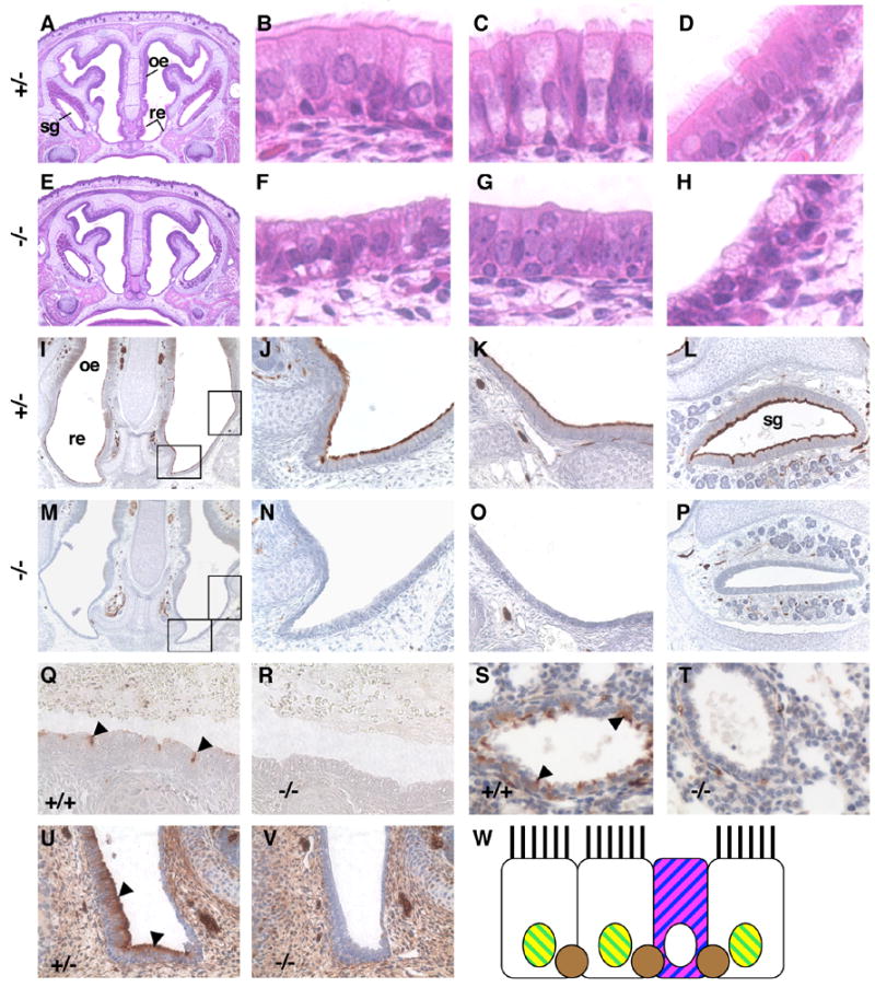
E2f4 mutant embryos lack ciliated cells in the airway epithelium. (A–H) Hematoxylin and eosin stained sections from E2f4+/− (A–D) and E2f4 mutant (E–H) 18.5 dpc littermate embryos. Panels A and E, coronal sections (original magnification ×4) showing the respiratory epithelium (re), olfactory epithelium (oe) and paranasal sinus serous gland (sg). High power images (original magnification ×40) of the respiratory epithelium (B, F), serous gland epithelium (C, G) and nasopharynx epithelium (D, H). (I–V) Acetylated α-tubulin, a component of cilia, was visualized by immunohistochemistry (brown stain). Panels I and M, coronal sections (original magnification ×10) with rectangles showing positions of the two adjacent panels. No staining was observed in the epithelium of E2f4 mutant embryos (M–O) compared with control embryos (I–K) (original magnification ×40) at 18.5 dpc. This phenotype was also observed in the serous gland (L, P) (original magnification ×20), in the trachea (Q, R) and bronchioles (S, T) at 18.5 dpc and in the nasal respiratory epithelium at 16.5 dpc (U, V) (original magnification ×40). Control genotypes are indicated (Q, S and U), E2f4−/− (R, T and V). Note that acetylated α-tubulin is also detected in neurons. Arrowheads point to ciliated cells in Q, S and U. W, Schematic of wild-type nasal respiratory epithelium with cell specific markers. Ciliated cells express Foxj1 (yellow) and Foxa1 (green). Goblet cell mucins stain with PAS (magenta) and Alcian Blue (blue). Basal cells express p63 (brown).