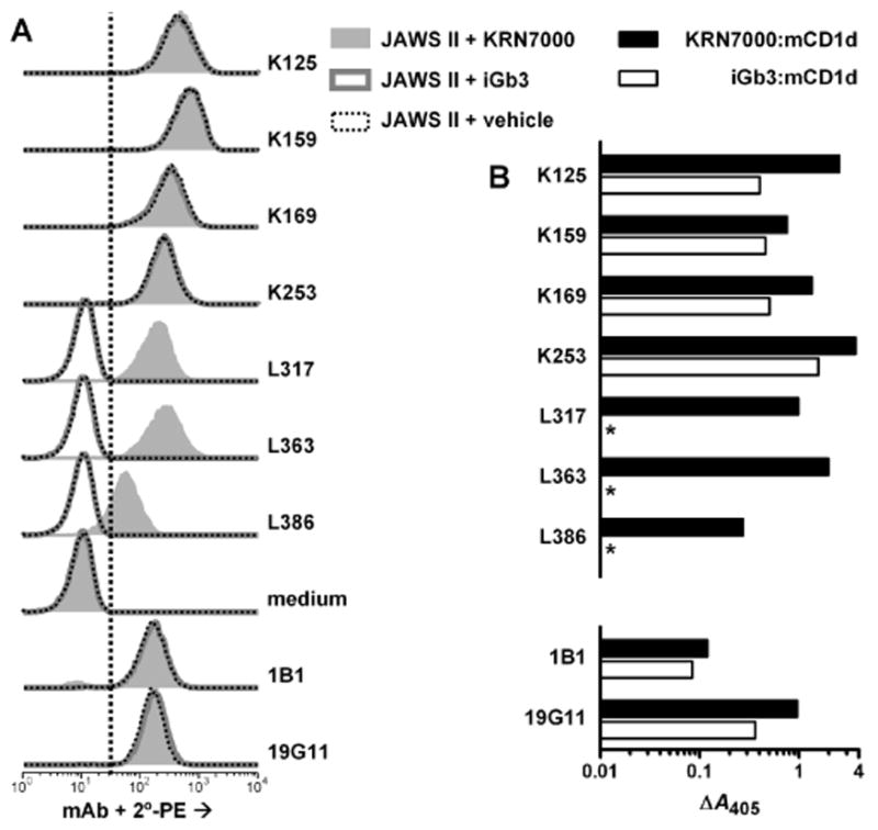
Figure 3.

Reactivity of hybridomas from immunized BALB/c and CD1d−/− mice. Seven mAb culture supernatants were assayed by FACS on JAWS II cells (A) or by ELISA against plate bound mCD1d protein (B) as in Fig. 2. These supernatants were from confluent cultures of twice-subcloned hybridomas derived from either CD1d−/− mouse #1 (K125, K159, K169 and K253) or from BALB/c mouse #3 (L317, L363, L386). Rat mAbs 1B1 and 19G11, used at 1 μg/ml and detected with rat-specific reagents, were included for comparison. *, specific A405 < 0.010. For (A), vertical dotted line indicates upper limit for negative control staining, as described in Figure 2. Negative control staining was deterimined here by the sample incubated without primary mAb, followed by PE-conjugated secondary antibody (“medium”). Previous experiments showed that no signals above this background level were observed when a nonbinding control IgG1 mAb (P3X63Ag8), whole normal mouse serum or normal rat polyclonal IgG were used under these staining conditions, and similar results were obtained for staining of RMA-S.mCD1d cells (data not shown).
