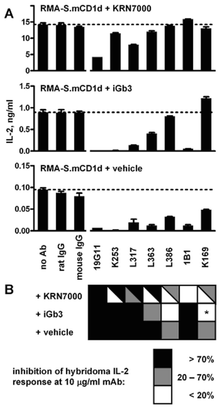
Figure 6.

mAb inhibition of iNKT cell hybridoma recognition of cell surface mCD1d. IL-2 production by the iNKT cell hybridoma DN3A4-1.2 was measured after incubation with KRN7000-, iGb3-, or vehicle-loaded mCD1d+ cells, in the presence of various mAbs at 10 μg/ml. (A) one representative experiment, with RMA-S.mCD1d cells. Mean ± SE of IL-2 levels from duplicate wells shown. Horizontal dotted lines indicate the amount of IL-2 obtained in the absence of added Ab. (B) checkerboard summary of data from four experiments, using either RMA-S.mCD1d or JAWS II dendritic cells. Darker boxes indicate more inhibition by a particular mAb of the IL-2 response to mCD1d+ cells loaded with each lipid. Split boxes show the range of results obtained. *, mAb K169 consistently enhanced hybridoma responses to iGb3-loaded mCD1d+ cells.
