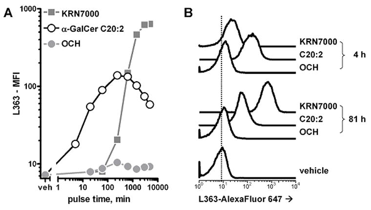
Figure 8.

Binding of mAb L363 to cells loaded in vitro with various α-GalCers. JAWS II dendritic cells were loaded with 100 nM KRN7000, α-GalCer C20:2 or OCH for various times at 37°C, after which cells were collected and stained with AlexaFluor 647-labeled mAb L363. mAb staining was detected by FACS. (A) median fluorescence intensity graphed against lipid antigen pulse time. Note that both axes are displayed on logarithmic scales. (B) FACS histograms for 4- and 81-hour loaded cells. The vertical dotted line is for comparison of the histograms with the readout from vehicle-loaded cells. Similar results were obtained with RMA-S.mCD1d cells.
