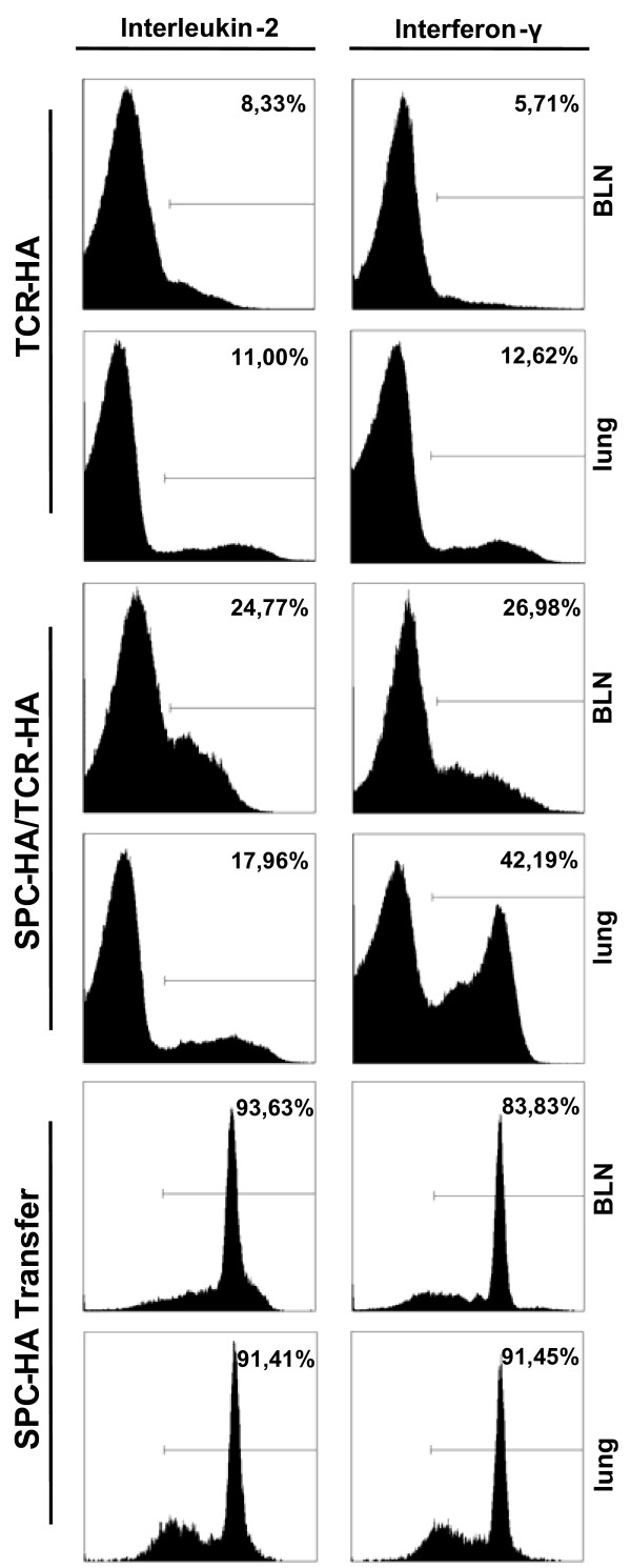
Figure 2.
Intracellular cytokine staining in CD4+ T cells. CD4+ T cells from the lung or bronchial lymph nodes (BLN) from either TCR-HA control mice, SPC-HA/TCR-HA double transgenic mice or SPC-HA mice adoptively transferred with HA-specific CD4+ T cells were analyzed by FACS for the expression of interleukin 2 and interferon γ.

