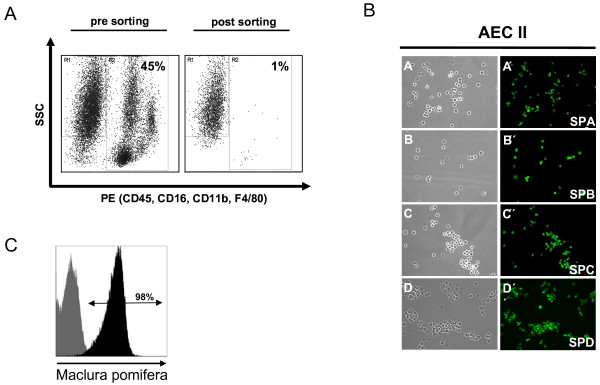
Figure 3.
Purification of alveolar type II epithelial cells by fluorescence-activated cell sorting. (A) Cell suspension obtained by enzymatic tissue disintegration and subsequent sequential filtration was labelled with antibodies to CD45, CD16, CD32, CD11b, and F4/80. Antibody negative AEC II were further distinguished from other cells by size and granularity. Reanalysis of sorted cells demonstrated an extremely low frequency of contaminating hematopoetic cells. (B) Sorted cells express surfactant proteins A, B, C and D. Cytospins of sorted AEC II cells were stained for the surfactant proteins A, B, C and D. Almost all cells were found to be positive for all four surfactant proteins. A, B, C and D represent phase contrast microscopy, A', B', C', and D' represent immunohistochemical stainings for the corresponding surfactant protein. (C) Staining of sorted AEC II with Maclura pomifera lectin revealed high purity of isolated cells. Black histogram indicates staining with the lectin, grey histogram indicates unstained cells.

