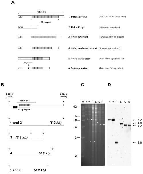
Figure 1. Generation of MHV-68 mutants.
A) Schematic presentation of viral mutants. B) Scheme of the expected fragments after digestion of viral DNA with the restriction enzyme EcoRI. Digestion of DNA from both parental virus and the 40 bp revertant with EcoRI results in a 5.2 kb wildtype fragment. Deletion of the 40 bp repeat results in the loss of the 5.2 kb fragment and in the generation of a new 2.8 kb fragment. Partial loss of repeat units results in a shift of the 5.2 kb fragment to 4.6 kb and 4.2 kb fragments, respectively. “P” indicates the probe used for Southern blot analysis, corresponding to nucleotides 25889-26711. C) Structural analysis of reconstituted virus genomes by ethidium bromide-stained agarose gel analysis of viral DNA digested with EcoRI. Lane 1: Parental virus; Lane 2: 40 bp revertant; Lane 3: Delta 40 bp mutant; Lane 4: 40 bp moderate mutant; Lane 5: 40 bp low mutant; Lane 6: M6STOP mutant. D) Southern blot analysis of the gel shown in panel C using probe “P” indicated in panel B. The expected fragments are indicated by dots (panel C) or by arrows (panel D). Marker (M) sizes (in kilobase pairs) are indicated on the left.

