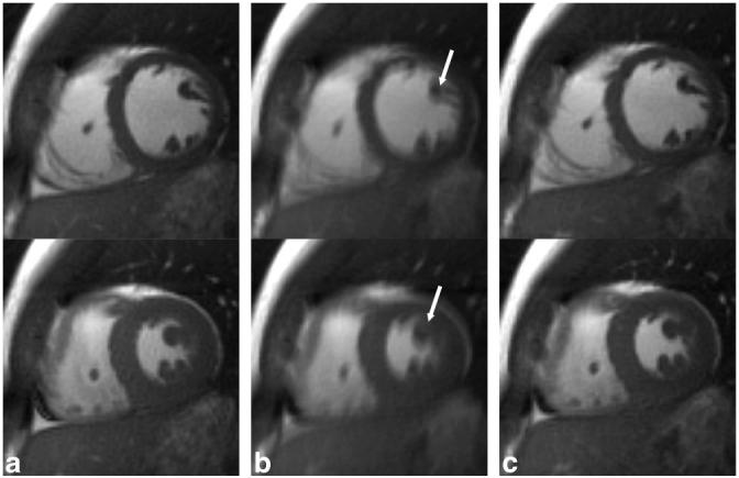
FIG. 5.

Representative end-diastole (top row) and end-systole (bottom row) short-axis images from a single volunteer reconstructed from a breath-hold acquisition (a) and a free-breathing acquisition with k-space averaging (b) as well as respiratory self-gating (c). Notice the blurring of fine anatomic structures in (b), arrows, and the relatively indistinguishable image quality between (a) and (c).
