
FIG. 1.
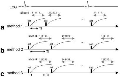
Timing for various acquisition methods: (a) method 1 using single slice per saturation preparation with short TI, (b) method 2 using single slice per saturation preparation with longer TI for flatter response and increased T1 contrast, and (c) method 3 using shot-to-shot slice interleaved acquisition with two slices acquired per saturation preparation and longer TI. Method 3 provides twice the spatial coverage of method 2 using the same TI. Method 3 uses TSENSE acceleration to reduce the imaging time for each pair of slices; therefore, the methods all have the same imaging window.