
Figure 1.
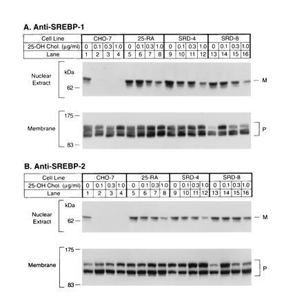
Immunoblot analysis of SREBP-1 (A) and SREBP-2 (B) in wild-type CHO-7 cells and mutant 25-hydroxycholesterol-resistant cells. On day 0, the indicated cell line was set up for experiments as described. On day 2, cells were switched to medium A containing 5% newborn calf lipoprotein-deficient serum, 50 μM sodium mevalonate, and 50 μM compactin supplemented with the indicated concentration of 25-hydroxycholesterol. After incubation for 16 h, the cells were harvested and fractionated as described, and aliquots of the nuclear extract (50 μg protein) and membrane fractions (100 μg protein) were subjected to SDS/PAGE. Immunoblot analysis was carried out with 30 μg/ml of IgG-2A4 (A) or 5 μg/ml IgG-7D4 (B) as described. Filters were exposed to film for 30 sec (A Upper) or 5 sec (A Lower and B). P and M denote the precursor and mature forms of SREBP-1 or SREBP-2, respectively.