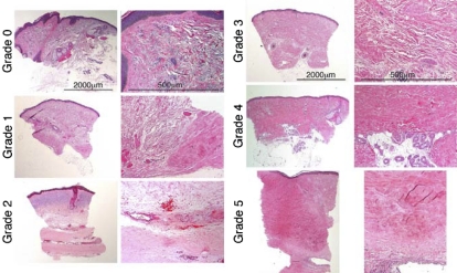
Figure 1.
Dermal fibrosis grades 0 to 5. Each pair represents a different dermal fibrosis grade. The left-hand column of each pair containing low-power views (original magnification, × 2.5), illustrates the spectrum of change in the overall dermal breadth and distribution of fibrosis. The right-hand column views for each pair (original magnification, × 10), illustrate the qualitative size, shape, distribution, and density in the dermal collagen bundles and surrounding extracellular matrix. All sections are stained with H&E. Grade 0 is thin curlicue collagen bundles with only minimal focal papillary dermal sclerosis. Grade 1 is less than 25% sclerosis with focal dense deep dermal fibrosis. In grade 2, the dense zone of fibrosis within the hypodermis comprises less than 50% of the overall dermal breadth. The remaining reticular dermis is composed of fine collagen bundles with actinic change (blue) in the upper dermis. Higher power shows the distinction between the sclerotic and normal collagen bundles. Grade 3 is extensive sclerosis—more than 50%—throughout the dermis with thickened collagen bundles admixed with thinner bundles and intervening extracellular matrix. Grade 4 is pandermal fibrosis with loss of most extracellular matrix without extension into hypodermis (or entrapment of eccrine coils) or surrounding fat. Grade 5 is markedly fibrotic and thickened—more than 4 mm—dermis replaced with dense waxy collagen with deep extension into hypodermis with fibrous incorporation of a muscular artery. See “Patients and methods; Assessment of skin biopsies” for more information on images.

