
Fig. 1.
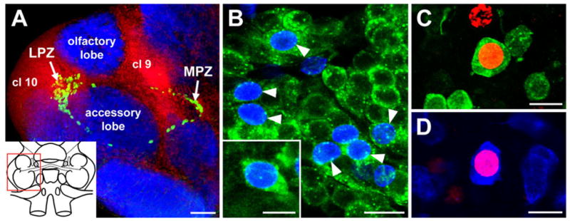
Neurogenesis in the olfactory pathway of the adult crayfish brain. A: Confocal image of the olfactory and accessory lobes of P. clarkii in a brain labelled immunocytochemically for BrdU (green) and Drosophila synapsin (blue) and counterstained with propidium iodide (red), a marker of nucleic acids. BrdU-labelled cells can be observed within both the lateral (LPZ) and medial (MPZ) proliferation zones. The inset shows a schematic drawing of the brain, and the red box delineates the region shown. B: BrdU (blue) and SIFamide (green) immunolabelling in cluster 10 six months after the exposure of the animal to BrdU. Double-labelled cells are indicated by the arrowheads. The inset shows a higher magnification image of the soma of a double-labelled neuron. C: Double labelling of a soma for BrdU (red) and orcokinin (green) in cluster 9 of an animal exposed 6 months previously to BrdU. D: Double labelling of a cluster 9 soma for BrdU (red) and allatostatin-like peptide (blue). cl 9, Soma cluster 9; cl 10, soma cluster 10; LPZ, lateral proliferation zone; MPZ, medial proliferation zone. Scale bars = 100 μm in A; 20 μm in B; 10 μm in inset in B; 15 μm in C,D.