
Fig. 11.
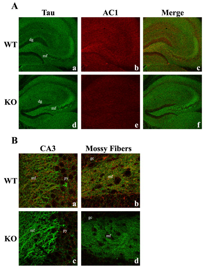
Immunohistochemical co-localization of AC1 protein with Tau in the adult mouse brain. A 100X confocal image of adult hippocampal sections demonstrates the specificity of the microtubule protein (extrasynaptic), Tau (green, Aa), and AC1 (red, Ab) and their co-localization (yellow, Ac), indicating the association of AC1 with extrasynaptic axonal proteins. In contrast, mice lacking both AC1 and AC8 exhibit only Tau immunoreactivity (Ad, Ae, Af). Further analysis at 900X demonstrates the co-localization of Tau and AC1 in hippocampal substructures such as the mossy fiber (mf)/CA3 pyramidal cell (py) junction (Ba) and the proximal mossy fiber pathway in the dentate hilus (Bb). dg, dentate gyrus; gc, dentate granule cells.