
Fig. 4.
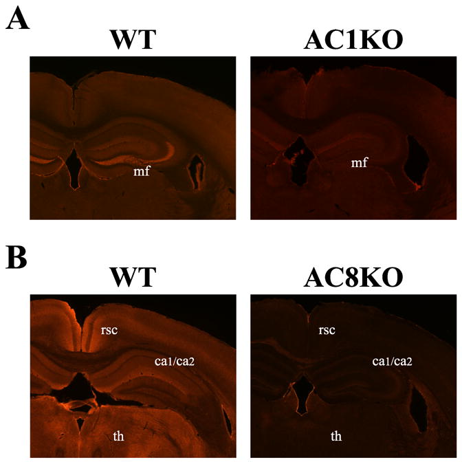
Localization of AC1 and AC8 protein in the mouse brain by immunohistochemistry. Representative coronal sections at 200X magnification from the adult mouse brain demonstrate unique patterns of protein distribution of AC1 and AC8. (A) AC1 immunoreactivity was robustly expressed in mossy fiber (mf) projections from the dentate hilus region extending to the stratum lucidum of the CA3 region of the hippocampus, with diffuse expression in the cortex and thalamus in the WT mouse. AC1 immunoreactivity is not detected in AC1KO mice demonstrating specific antigen recognition. (B) AC8 immunoreactivity is abundantly present in the CA1/CA2 (ca1/ca2) region in the hippocampus, habenula, retrosplenial cortex (rsc) and various thalamic nuclei (th) with weaker expression in the cerebral cortex in the WT mouse. AC8 immunoreactivity is not detected in AC8KO mice.