
Fig. 7.
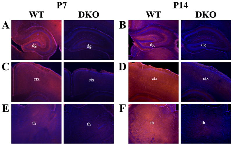
Localization of AC1 protein in the developing mouse brain by immunohistochemistry. Representative coronal sections at 400X magnification from the neonatal mouse brain at postnatal day 7 (P7) and postnatal day 14 (P14) illustrate the dynamic progression of AC1 protein expression. (A) AC1 protein is widely expressed throughout the hippocampal formation at P7. (B) Expression of AC1 resembles adult expression by P14, with specific staining localized to the mossy fiber projections and the dendritic arbors of the dentate gyrus (dg). Cortical (ctx) expression of AC1 is diffuse at both (C) P7 and (D) P14, without localization to a particular cell layer. AC1 protein is expressed widely throughout various thalamic (th) nuclei at both (E) P7 and (F) P14. Sections are counterstained with DAPI (blue) to provide anatomical context.